LDS Sample Loading Buffer (4X): A Comprehensive Guide

LDS (Lithium Dodecyl Sulfate) Sample Loading Buffer (4X) is an essential reagent used in molecular biology, particularly in protein electrophoresis applications such as SDS-PAGE (sodium dodecyl sulfate-polyacrylamide gel electrophoresis). It is a key component for preparing protein samples for gel electrophoresis, ensuring efficient protein denaturation and accurate molecular weight determination.

Composition and Function

LDS Sample Loading Buffer contains several important components that facilitate protein sample preparation:

  • Lithium Dodecyl Sulfate (LDS): A detergent that helps solubilize proteins and maintain their denatured state. Unlike SDS, LDS is often preferred for certain applications due to its ability to solubilize hydrophobic proteins more effectively.
  • Reducing Agents (DTT or β-mercaptoethanol): These agents break disulfide bonds, ensuring that proteins are fully denatured and linearized before electrophoresis.
  • Tracking Dyes (Bromophenol Blue or Coomassie G250): These dyes help track the progress of electrophoresis by providing visible markers during gel running.
  • Buffering Agents: Maintain the pH stability of the solution to ensure optimal protein denaturation and migration.
  • Glycerol: Increases the density of the sample, allowing it to settle into the gel wells properly without diffusing away.

Preparation and Usage

The LDS Sample Loading Buffer (4X) is typically used by diluting it to 1X concentration in the final sample preparation. The following steps outline the standard protocol for using LDS Sample Loading Buffer:

  1. Sample Preparation: Mix protein samples with LDS Sample Loading Buffer at a 1:3 ratio (e.g., 10 µL of protein sample with 30 µL of 4X buffer).
  2. Addition of Reducing Agent: If reducing conditions are required, add 1/10 volume of 10X reducing agent such as DTT or β-mercaptoethanol.
  3. Denaturation: Heat the mixture at 70°C for 10 minutes to ensure complete protein denaturation.
  4. Loading onto the Gel: Once the sample has cooled, it can be loaded onto an SDS-PAGE gel for electrophoresis.

For more details on the preparation of LDS Sample Loading Buffer, visit the National Institutes of Health (NIH) website (nih.gov).

Applications in Research

LDS Sample Loading Buffer is widely used in various research applications, including:

1. Western Blotting

Western blotting is a widely used technique for detecting specific proteins in a sample. LDS buffer ensures that proteins are fully denatured and separated by molecular weight before being transferred to a membrane for antibody-based detection. More details on Western blotting can be found at the National Cancer Institute (NCI) (cancer.gov).

2. Protein Purification and Quality Control

LDS Sample Loading Buffer is frequently used in protein purification workflows to assess protein purity and molecular weight. Laboratories use SDS-PAGE to analyze purified proteins before downstream applications such as mass spectrometry. The Frederick National Laboratory for Cancer Research provides useful protocols for protein analysis (frederick.cancer.gov).

3. Structural and Functional Protein Studies

Scientists studying protein folding and interactions use LDS Sample Loading Buffer to prepare proteins for electrophoresis-based structural analyses. Institutions such as the National Center for Biotechnology Information (NCBI) (ncbi.nlm.nih.gov) offer valuable resources on protein studies.

Advantages of LDS Over SDS Loading Buffers

Compared to traditional SDS buffers, LDS Sample Loading Buffer offers several advantages:

  • Better solubilization of membrane proteins: LDS is more effective at solubilizing hydrophobic proteins, making it ideal for studying membrane proteins.
  • Improved protein separation: LDS buffer provides sharper bands and better resolution, which is particularly beneficial when analyzing low-molecular-weight proteins.
  • Compatibility with alternative gel chemistries: LDS is often used in Bis-Tris gels instead of traditional Tris-glycine gels, leading to better separation and reproducibility.

For a comparative analysis of different loading buffers, visit the National Institute of Standards and Technology (NIST) (nist.gov).

Best Practices and Considerations

1. Proper Storage and Handling

LDS Sample Loading Buffer should be stored at -20°C for long-term storage. If using frequently, it can be kept at 4°C for short-term use. Repeated freeze-thaw cycles should be avoided to prevent buffer degradation.

2. Optimization for Specific Applications

Depending on the type of gel and electrophoresis conditions used, the composition of the buffer may need to be adjusted. Some proteins require additional reducing agents or alternative detergents to optimize solubility.

For guidance on optimizing electrophoresis conditions, the National Science Foundation (NSF) (nsf.gov) provides valuable insights.

Alternative Sample Buffers

While LDS Sample Loading Buffer is widely used, researchers may sometimes opt for alternative loading buffers such as:

  • Laemmli Buffer (SDS-based): Traditionally used in Tris-glycine gels.
  • Native Loading Buffers: Used when studying protein interactions that require non-denaturing conditions.
  • Urea-based Buffers: Used for proteins that aggregate in traditional SDS or LDS buffers.

More information on electrophoresis buffer formulations can be found at PubMed Central (PMC) (ncbi.nlm.nih.gov/pmc).

Conclusion

LDS Sample Loading Buffer (4X) is a fundamental reagent in protein electrophoresis, offering superior solubilization, sharp protein bands, and compatibility with various gel systems. Its widespread applications in research, from Western blotting to protein purification, make it a staple in molecular biology laboratories. Understanding its composition, usage, and advantages can help researchers optimize their protein electrophoresis workflows.

For further reading on LDS Sample Loading Buffer and related protocols, visit official government and educational sites such as:


 

VAHTS Dual UMI UDB Adapters Set 7 for MGI: A Breakthrough in High-Throughput Sequencing

Introduction

The VAHTS Dual UMI UDB Adapters Set 7 for MGI is a state-of-the-art library preparation solution designed for next-generation sequencing (NGS) platforms. This adapter set incorporates unique molecular identifiers (UMIs) to enhance sequencing accuracy and minimize errors caused by PCR duplication. These adapters are specifically optimized for MGI sequencing platforms, providing researchers with a robust tool for genomics applications, including cancer research, transcriptomics, and metagenomics.

Importance of Unique Molecular Identifiers (UMIs) in NGS

UMIs are short, random sequences incorporated into DNA fragments before PCR amplification. They serve as molecular barcodes, allowing researchers to distinguish between true biological variants and sequencing errors. The VAHTS Dual UMI UDB Adapters Set 7 ensures:

  • Error Correction: Eliminates PCR and sequencing-induced errors (NIH).
  • Improved Quantification: Reduces amplification bias, providing more accurate transcript quantification (NCBI).
  • High Sensitivity: Enables the detection of low-frequency mutations in applications such as liquid biopsy (FDA).

Key Features of VAHTS Dual UMI UDB Adapters Set 7

This adapter set is specifically designed to work with MGI sequencing platforms, offering multiple advantages:

  • Dual UMI System: Enhances error correction and deduplication (NIH NCBI).
  • Optimized for MGI Platforms: Provides high compatibility with MGI’s DNBSEQ technology (MGI Tech).
  • High-Throughput Applications: Ideal for RNA sequencing, whole-genome sequencing, and targeted sequencing (CDC).
  • Efficient Sample Indexing: Enables multiplexing of large sample sets with minimal cross-contamination (WHO).

Applications of VAHTS Dual UMI UDB Adapters

1. Cancer Genomics

Ultra-sensitive detection of mutations is crucial in oncology. This adapter set allows for precise variant calling in tumor sequencing projects (National Cancer Institute).

2. Transcriptomics and Gene Expression Studies

With accurate UMI-based quantification, researchers can analyze RNA expression profiles with reduced technical noise (NIH Gene Expression Omnibus).

3. Metagenomics and Microbial Diversity Studies

UMI-based error correction is essential in metagenomics applications, ensuring accurate representation of microbial communities (CDC Pathogen Genomics).

4. Clinical and Translational Research

Accurate sequencing is vital for developing personalized medicine approaches. This adapter set ensures reliable genomic data for clinical applications (FDA Precision Medicine).

Quality Control and Regulatory Compliance

To ensure optimal performance, laboratories must adhere to stringent quality control measures and regulatory standards:

  • U.S. Food and Drug Administration (FDA) – Sets guidelines for NGS-based clinical applications (FDA NGS Guidelines).
  • World Health Organization (WHO) – Establishes best practices for genomic sequencing and pathogen surveillance (WHO Genomic Sequencing).
  • Centers for Disease Control and Prevention (CDC) – Provides recommendations for implementing NGS in public health laboratories (CDC Genomic Testing).

Challenges in UMI-Based Sequencing

Despite its advantages, UMI-based sequencing presents some challenges:

  • Data Analysis Complexity: UMI deduplication requires advanced bioinformatics pipelines (NCBI Genomics).
  • Library Preparation Optimization: Ensuring efficiency in UMI incorporation is critical for accurate sequencing results (NIH Research).
  • Cost Considerations: While improving accuracy, UMI-based approaches may require additional computational resources and reagent costs (FDA Genomics Research).

Future Directions and Innovations

As sequencing technology advances, the integration of UMIs will become more widespread in genomics research. Emerging trends include:

  • AI-Driven Sequencing Error Correction: Machine learning approaches to refine UMI deduplication (NIH AI in Genomics).
  • Expansion of Long-Read Sequencing: Combining UMIs with long-read sequencing for improved structural variant detection (WHO Long-Read Genomics).
  • Automated High-Throughput Library Prep: Robotics and automation will enhance efficiency in sample preparation (CDC Genomic Surveillance).

Conclusion

The VAHTS Dual UMI UDB Adapters Set 7 for MGI represents a significant advancement in high-throughput sequencing. By integrating dual unique molecular identifiers (UMIs), these adapters enhance sequencing accuracy, reduce errors, and improve variant detection. With its broad applications in cancer research, transcriptomics, metagenomics, and clinical genomics, this technology plays a crucial role in modern genomics. Adhering to FDA, WHO, and CDC guidelines ensures the quality and reliability of sequencing data, making this adapter set a valuable tool for researchers worldwide.

For further information, visit:

Extra Murine Hepatitis Virus (MHV) PCR Qualitative Quality Control

Introduction

Murine Hepatitis Virus (MHV) is a coronavirus that primarily infects mice, causing significant research implications in laboratory animal studies. As an essential model for understanding coronaviruses, MHV requires precise detection methods, with PCR (Polymerase Chain Reaction) qualitative testing being the gold standard. Ensuring accurate and reliable MHV PCR qualitative testing necessitates rigorous quality control measures to validate test results and maintain laboratory accuracy.

Importance of MHV Detection in Research

MHV is widely studied due to its structural and genetic similarities to human coronaviruses, including SARS-CoV and SARS-CoV-2. This virus plays a critical role in virology and immunology research, making MHV PCR qualitative testing indispensable for maintaining pathogen-free laboratory environments (NIH, NCBI).

Key Reasons for MHV Detection:

  • Animal Health Monitoring: Prevents virus spread in research mouse colonies (CDC).
  • Coronavirus Research: Provides insights into pathogenesis, immunity, and therapeutic responses (NCBI).
  • Standardized Lab Practices: Maintains experimental reproducibility (FDA).
  • Biosafety Compliance: Ensures proper containment of pathogens in lab settings (WHO).

PCR-Based Detection of MHV

PCR is the most sensitive and specific technique for detecting MHV, distinguishing between infected and non-infected mice with high accuracy (PubMed). The qualitative PCR test confirms the presence or absence of viral RNA, making it essential for maintaining virus-free lab environments.

Advantages of PCR for MHV Detection

  • High Sensitivity & Specificity: Detects low viral loads accurately (FDA).
  • Rapid Turnaround Time: Provides results within hours (CDC).
  • Minimal Contamination Risk: Uses automated, closed-system PCR platforms (WHO).
  • Reliable Quality Control Measures: Ensures test reproducibility (NIH).

Quality Control in MHV PCR Testing

Effective quality control (QC) ensures that MHV PCR tests produce reliable and reproducible results. Laboratories must adhere to standardized protocols established by global health authorities.

Essential Quality Control Measures:

  1. Positive and Negative Controls – Ensures accuracy by running known positive and negative samples alongside test samples (CLSI).
  2. Standardized Sample Handling – Reduces pre-analytical errors through proper sample collection and storage (WHO).
  3. Reagent and Kit Validation – Regularly verifies the effectiveness of PCR reagents (FDA).
  4. Cross-Contamination Prevention – Implements strict laboratory workflows to minimize contamination risks (CDC).
  5. Regular Equipment Calibration – Ensures PCR machines provide consistent and accurate results (NIH).

Applications of MHV PCR Quality Control in Research

1. Prevention of False Positives and Negatives

QC measures help differentiate true infections from sample contamination, preventing misleading results in critical studies (FDA).

2. Ensuring Reproducibility in Studies

High-quality control standards maintain consistent results across different laboratories, ensuring reproducibility (WHO).

3. Biosafety and Laboratory Compliance

MHV research must adhere to biosafety guidelines to prevent accidental viral outbreaks in laboratory settings (CDC).

4. Optimization of Research Models

MHV-free mouse models are essential for highly controlled immunology and virology research (NIH).

Regulatory Guidelines for MHV PCR Quality Control

To ensure accurate MHV testing, laboratories follow strict regulatory frameworks provided by global health agencies.

Challenges in MHV PCR Quality Control

Despite advancements in PCR technology, maintaining quality control presents challenges:

  • Inter-Laboratory Variability: Differences in PCR methodologies can lead to inconsistent results (WHO).
  • Risk of Sample Contamination: Requires stringent protocols to avoid false positives (CDC).
  • Supply Chain Issues: Shortages of high-quality PCR reagents affect test accuracy (FDA).

Future Directions in MHV PCR Testing

The future of MHV PCR testing involves integrating next-generation sequencing (NGS) and digital PCR (dPCR) to enhance detection accuracy. Automated sample processing and artificial intelligence-driven quality control are also emerging trends (NIH).

Conclusion

MHV PCR Qualitative Quality Control is essential for ensuring accurate, reliable, and standardized testing in research and laboratory settings. Adhering to WHO, CDC, FDA, and CLSI guidelines guarantees high-quality results, ultimately improving the reproducibility and credibility of MHV-related studies. As technology advances, incorporating automated quality control measures and digital molecular diagnostics will further enhance the field of virology research.

For additional resources, visit:


Cytosolic Non-Specific Dipeptidase ELISA (CNDP2): A Critical Tool for Protein Quantification

Introduction

Cytosolic non-specific dipeptidase (CNDP2) is a crucial enzyme involved in intracellular protein metabolism and amino acid recycling. CNDP2 is widely expressed in human tissues and plays an essential role in various physiological processes, including peptide degradation and metabolic regulation. The ELISA (Enzyme-Linked Immunosorbent Assay) for CNDP2 provides researchers and clinicians with a powerful tool to quantitatively measure this enzyme’s presence in biological samples. This article explores the importance of CNDP2, the advantages of using ELISA, and regulatory considerations in protein quantification.


Biological Role of CNDP2

CNDP2, also known as Carnosine Dipeptidase 2, is a member of the M20 family of metalloproteases (NCBI). This enzyme primarily functions to hydrolyze dipeptides in the cytosol, contributing to amino acid metabolism and cellular homeostasis (PubMed).

CNDP2 in Metabolic Processes

  • Protein and Peptide Degradation: CNDP2 catalyzes the hydrolysis of various dipeptides, playing a significant role in amino acid recycling (Protein Data Bank).
  • Antioxidant Regulation: The enzyme contributes to carnosine metabolism, which has antioxidant properties and potential neuroprotective effects (NIH).
  • Cancer and Disease Association: CNDP2 has been implicated in cancer metabolism and neurological disorders (National Cancer Institute).

The Importance of CNDP2 ELISA

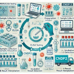
ELISA is a widely used technique for detecting and quantifying proteins in biological samples. It is especially useful in research and clinical diagnostics due to its high specificity and sensitivity. CNDP2 ELISA provides valuable insights into enzyme activity and disease progression in different physiological and pathological contexts.

Advantages of ELISA in CNDP2 Detection

  • High Sensitivity & Specificity: ELISA can detect CNDP2 at nanogram-level concentrations, ensuring precise quantification (FDA).
  • Rapid and Reliable: The assay provides quick results with minimal sample preparation (CDC).
  • Standardized Protocols: Ensures reproducibility across different laboratories (WHO).
  • Cost-Effective: Compared to mass spectrometry and Western blotting, ELISA is more accessible for large-scale studies (CLSI).

Applications of CNDP2 ELISA in Research and Medicine

1. Biomedical Research

Researchers utilize CNDP2 ELISA to study the enzyme’s role in various biological pathways and diseases. This method is widely used to monitor expression levels in cancer, metabolic syndromes, and neurological disorders (NIH).

2. Disease Biomarker Identification

CNDP2 expression has been linked to cancer progression, making it a potential biomarker for disease detection and prognosis (PubMed).

3. Pharmaceutical and Drug Development

ELISA assays help screen potential inhibitors of CNDP2 activity, which could lead to new therapeutic strategies for metabolic and neurological disorders (FDA Drug Development).

4. Clinical Diagnostics

Quantifying CNDP2 levels in patient samples assists in disease diagnosis and monitoring of treatment responses (CDC Laboratory Standards).

Regulatory Considerations for CNDP2 ELISA

To ensure accurate and reproducible results, laboratories must comply with established quality control and regulatory standards.

  • World Health Organization (WHO) – Provides guidelines for diagnostic assay validation (WHO Laboratory Guidance).
  • Clinical and Laboratory Standards Institute (CLSI) – Establishes best practices for immunoassay development (CLSI Molecular Guidelines).
  • U.S. Food and Drug Administration (FDA) – Regulates ELISA-based diagnostic kits for clinical use (FDA IVD Regulations).
  • Centers for Disease Control and Prevention (CDC) – Provides laboratory quality control measures to enhance assay performance (CDC Laboratory Quality).

Challenges in CNDP2 Quantification

Despite the advantages of ELISA, some challenges remain in CNDP2 measurement:

  • Sample Variability: Differences in biological samples can affect enzyme activity and concentration (WHO Laboratory Testing).
  • Cross-Reactivity: Some antibodies may react with structurally similar proteins, leading to false-positive results (FDA Reference Materials).
  • Assay Standardization: Differences in assay kits and protocols can lead to variations in results, emphasizing the need for standardized quality control measures (CDC Lab Standards).

Future Directions and Innovations

Recent advancements in multiplex ELISA and digital immunoassays are improving the sensitivity and specificity of CNDP2 detection. Emerging technologies such as microfluidic ELISA platforms and automated high-throughput screening hold promise for enhancing diagnostic capabilities (NIH Advanced Molecular Testing).

Conclusion

The Cytosolic Non-Specific Dipeptidase (CNDP2) ELISA is a crucial tool for understanding protein metabolism and its role in disease. It provides a reliable and efficient method for measuring CNDP2 levels in research and clinical settings. Compliance with WHO, FDA, CLSI, and CDC guidelines ensures accurate, reproducible, and high-quality diagnostic results. As technology advances, the future of CNDP2 quantification promises even greater precision and clinical applicability.

For further information, visit:


Human Parvovirus B19 PCR Quantitative Quality Control L1: A Critical Tool in Diagnostic Virology

Introduction

Human Parvovirus B19 is a small, non-enveloped DNA virus that primarily causes erythema infectiosum (Fifth Disease) and is associated with more severe conditions such as aplastic anemia and hydrops fetalis. Accurate detection and quantification of B19 DNA are essential for clinical diagnosis, blood donor screening, and epidemiological studies. The PCR Quantitative Quality Control L1 is a critical component in ensuring the reliability and accuracy of molecular testing for this virus.

Human Parvovirus B19: Clinical Significance and Molecular Diagnosis

Human Parvovirus B19 primarily targets erythroid progenitor cells, leading to transient anemia, particularly in individuals with underlying hematologic conditions. Infection is particularly concerning in pregnant women and immunocompromised patients (CDC).

Polymerase Chain Reaction (PCR) is the gold standard for detecting B19 DNA due to its high sensitivity and specificity. Quantitative PCR (qPCR) allows for precise measurement of viral load, which is crucial for monitoring infection progression and treatment response (NIH).

Importance of PCR Quantitative Quality Control L1

The PCR Quantitative Quality Control L1 is a standardized reference material used in laboratory quality assurance programs. It ensures consistency in PCR assays by monitoring assay performance, detecting potential contamination, and validating results (WHO).

Key Functions of Quality Control L1:

  • Assay Validation: Ensures the accuracy and precision of qPCR-based B19 detection (FDA).
  • Reproducibility: Helps standardize results across different laboratories and diagnostic platforms (CLSI).
  • Contamination Monitoring: Detects potential contamination or cross-reactivity in PCR assays (CDC).
  • Performance Tracking: Evaluates test sensitivity and specificity over time (CAP).

Standardized Testing Procedures and Regulatory Guidelines

To ensure high-quality diagnostic testing, laboratories must adhere to international guidelines set by organizations such as:

  • World Health Organization (WHO) – Provides standards for virological testing and quality assurance (WHO Laboratory Guidance).
  • Clinical and Laboratory Standards Institute (CLSI) – Establishes best practices for molecular diagnostics (CLSI Molecular Guidelines).
  • U.S. Food and Drug Administration (FDA) – Regulates in vitro diagnostic tests, including qPCR assays (FDA IVD).

Applications of B19 PCR Quantitative Quality Control

1. Clinical Diagnostics

Quantitative PCR is widely used in diagnosing acute B19 infection, particularly in high-risk patients such as pregnant women and immunocompromised individuals. Quality Control L1 ensures the reliability of these critical tests (PubMed).

2. Blood Screening

Blood banks use qPCR to screen donations for B19 contamination, as the virus can be transmitted via blood products (AABB). Regular quality control assessments help maintain blood safety standards (CDC Blood Safety).

3. Epidemiological Surveillance

Tracking the prevalence of B19 in populations requires accurate molecular diagnostics. PCR quality control ensures that surveillance data are reliable and comparable across different study sites (WHO Epidemiology).

4. Vaccine and Therapeutic Research

Researchers studying antiviral therapies and vaccine development rely on standardized qPCR assays to evaluate treatment efficacy. Quality control standards are essential for producing reproducible and validated results (NIH Clinical Trials).

Challenges in B19 PCR Quality Control

Despite advancements in molecular diagnostics, challenges remain in ensuring high-quality results:

  • Variability Between Assay Platforms: Different PCR systems may yield varying results, underscoring the need for uniform quality control measures (CDC Laboratory Standards).
  • Contamination Risks: False positives due to laboratory contamination can compromise diagnostic accuracy (WHO Laboratory Safety).
  • Standard Reference Material Availability: Access to certified reference materials can be limited in certain regions, affecting test standardization (FDA Reference Materials).

Future Directions

Advancements in digital PCR (dPCR) and next-generation sequencing (NGS) are improving B19 detection accuracy. Implementing automated quality control measures can further enhance diagnostic reliability (NIH Advanced Molecular Testing).

Conclusion

Human Parvovirus B19 PCR Quantitative Quality Control L1 is essential for maintaining the accuracy, reliability, and standardization of molecular diagnostics. With its applications spanning clinical diagnostics, blood screening, epidemiology, and research, it plays a crucial role in disease management and public health. Compliance with regulatory guidelines from the WHO, FDA, CLSI, and CDC ensures high-quality testing and reliable patient outcomes.

For further information, visit CDC Parvovirus B19, FDA Laboratory Guidelines, and WHO Virology Standards.


Understanding the Human Proton Myo-Inositol Cotransporter (SLC2A13) and Its Role in Biomedical Research

Introduction

The human proton myo-inositol cotransporter (SLC2A13), also known as HMIT, is a crucial membrane transport protein that facilitates the uptake of myo-inositol, a key molecule involved in cell signaling and osmoregulation. This transporter is primarily expressed in the brain, where it plays a significant role in neural function and cognitive processes. Understanding the physiological and pathological implications of SLC2A13 is essential for advancing research in neurological disorders, including Alzheimer’s disease. In this article, we explore the function, structure, and diagnostic tools used to study SLC2A13, particularly the Enzyme-Linked Immunosorbent Assay (ELISA).

Structure and Function of SLC2A13

SLC2A13 is part of the glucose transporter (GLUT) family, specifically the solute carrier 2 family (SLC2). Unlike other glucose transporters, SLC2A13 operates as a proton-coupled transporter, relying on a proton gradient to facilitate myo-inositol uptake. This distinguishes it from passive glucose transporters such as GLUT1 or GLUT4 (UniProt).

Expression and Physiological Role

SLC2A13 is predominantly expressed in the brain, particularly in regions such as the cerebral cortex, hippocampus, and amygdala (The Human Protein Atlas). Its localization suggests a significant role in neural signal transduction, synaptic plasticity, and cognitive functions. Research indicates that myo-inositol transport is critical for maintaining phosphatidylinositol signaling pathways, which are involved in neurotransmission and neuroprotection.

SLC2A13 and Neurodegenerative Diseases

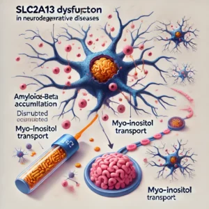
Recent studies have suggested that SLC2A13 is associated with Alzheimer’s disease due to its potential involvement in amyloid-beta production. Amyloid-beta accumulation is a hallmark of Alzheimer’s pathology, and disruptions in inositol metabolism have been linked to neurodegeneration (NCBI Gene).

Myo-inositol is a precursor for inositol phosphates and phosphatidylinositol, which are essential for maintaining cellular homeostasis. Dysregulation of inositol transport can lead to altered phosphoinositide signaling, which in turn may contribute to cognitive decline and neurodegeneration (PubMed).

The Importance of ELISA in SLC2A13 Research

Enzyme-Linked Immunosorbent Assay (ELISA) is a widely used analytical technique for detecting and quantifying proteins, including SLC2A13. This method allows researchers to measure SLC2A13 expression levels in various biological samples such as plasma, serum, and brain tissue extracts.

How ELISA Works

ELISA is based on the antigen-antibody reaction. Specific antibodies targeting SLC2A13 are immobilized on a microplate, allowing the capture and quantification of the target protein from biological samples. Detection is typically achieved through colorimetric or fluorometric readouts, providing high sensitivity and specificity (NIH Assay Guidance Manual).

Applications of ELISA in Neuroscience

Researchers use ELISA to study SLC2A13 expression in neurological disorders. By quantifying SLC2A13 levels in patient samples, scientists can investigate potential correlations between its expression and disease states such as Alzheimer’s or bipolar disorder (National Institute on Aging).

Several commercial ELISA kits are available for detecting SLC2A13. These kits typically offer high specificity and sensitivity, making them valuable tools for studying the protein’s role in health and disease (LSBio, MyBiosource).

Future Directions and Clinical Implications

Given the critical role of SLC2A13 in brain function, further research is needed to elucidate its involvement in neurodegenerative and psychiatric disorders. Advanced techniques such as single-cell RNA sequencing and proteomics may provide deeper insights into the molecular mechanisms regulated by SLC2A13 (Human Genome Research Institute).

Potential therapeutic strategies targeting SLC2A13 could involve modulating its expression or function to improve inositol metabolism in neurodegenerative diseases. For example, pharmacological agents that enhance myo-inositol uptake may hold promise for treating cognitive disorders (FDA Drug Development).

Conclusion

SLC2A13 is a key transporter involved in myo-inositol uptake, playing a crucial role in brain physiology and disease. Understanding its function and regulation is vital for developing novel therapeutic strategies for neurodegenerative disorders. ELISA-based detection methods offer a reliable approach for quantifying SLC2A13 levels, facilitating research into its potential as a biomarker and drug target. Continued studies on SLC2A13 could pave the way for new treatments for Alzheimer’s disease and other neurological conditions.

For further information, explore resources from the National Institutes of Health, Centers for Disease Control and Prevention, and PubMed.